
PISA geometry
| View | Zoomed colour Doppler of the regurgitant orifice, variance mapping off, baseline shifted in the direction of the jet. |
|---|---|
| Timing | Colour frame at the time of maximal regurgitant jet velocity by CW. |
| Caliper / trace | Radius from the first aliasing contour (abrupt colour change) to the vena contracta, along the centreline of the convergence. |
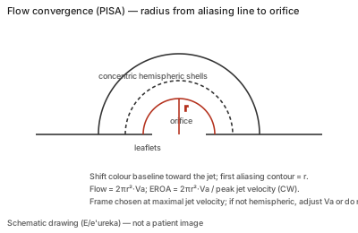
Correct example
Baseline shifted (down for MR into the LA on TTE) so that a well-defined hemisphere appears; radius measured to the orifice, with dark zones of perpendicular flow marking the orifice position; EROA = 2πr²·Va / peak velocity, RVol = EROA × VTI.
Common error
Measuring a flattened or constrained convergence as if hemispheric, taking the radius on a frame that does not match peak velocity, or forcing the method on an elongated functional orifice.
Effect on interpretation
The radius is squared, so geometry errors dominate; non-hemispheric or elongated convergence underestimates flow and EROA, and a conical orifice plane (TR, tenting) means the convergence covers more than a hemisphere. If a hemisphere cannot be obtained, the method is not reported.
Why
Flow accelerates toward the orifice in concentric isovelocity shells; colour aliasing exposes one shell whose surface area times its velocity is the instantaneous flow rate.