
LVOT diameter and sample position
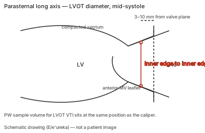
| View | Zoomed parasternal long axis showing the centre axis of the LVOT and the aortic cusp insertion points. |
|---|---|
| Timing | Mid-systole. |
| Caliper / trace | Inner edge to inner edge, compacted septum to anterior mitral leaflet, about 3–10 mm from the valve plane, at the same position as the PW sample volume used for the LVOT VTI. |
Correct example
Zoomed PLAX, gain adjusted to the blood–tissue interface, caliper placed perpendicular to the outflow tract 3–10 mm below the cusp insertions in mid-systole; the PW sample volume for the VTI is then placed at that same distance from the valve.
Common error
Measuring the aortic annulus (cusp insertion) or an oblique, non-zoomed image and pairing it with a VTI sampled at a different level; measuring a single beat in an irregular rhythm.
Effect on interpretation
The diameter is squared to obtain a circular area, so a small error in diameter is magnified in stroke volume and continuity AVA; underestimating the LVOT area is the most important source of AVA underestimation.
Why
Stroke volume is CSA × VTI, and CSA assumes a circle from the diameter; the diameter and the velocity sample must come from the same level of the outflow tract for the product to describe one flow.