
Chronic mitral regurgitation by Doppler echocardiography
Native valves
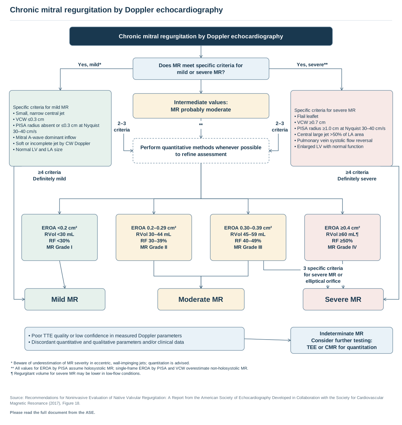
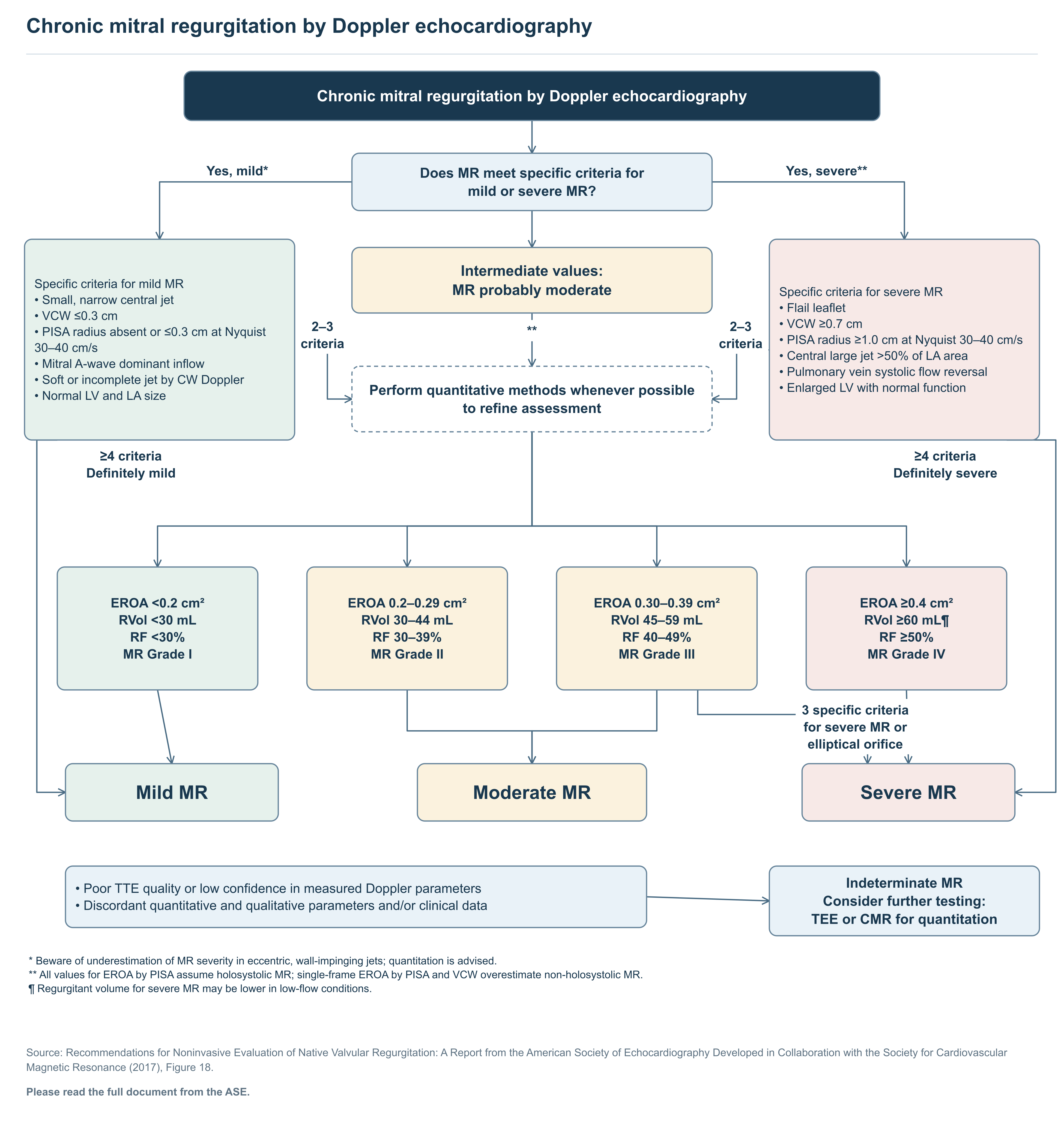
Text of this figure
- Chronic mitral regurgitation by Doppler echocardiography
- Yes, mild*
- Yes, severe**
- **
- 2–3
- criteria
- 2–3
- criteria
- ≥4 criteria
- Definitely mild
- ≥4 criteria
- Definitely severe
- 3 specific criteria
- for severe MR or
- elliptical orifice
- Chronic mitral regurgitation by Doppler echocardiography
- Does MR meet specific criteria for
- mild or severe MR?
- Specific criteria for mild MR
- • Small, narrow central jet
- • VCW ≤0.3 cm
- • PISA radius absent or ≤0.3 cm at Nyquist
- 30–40 cm/s
- • Mitral A-wave dominant inflow
- • Soft or incomplete jet by CW Doppler
- • Normal LV and LA size
- Specific criteria for severe MR
- • Flail leaflet
- • VCW ≥0.7 cm
- • PISA radius ≥1.0 cm at Nyquist 30–40 cm/s
- • Central large jet >50% of LA area
- • Pulmonary vein systolic flow reversal
- • Enlarged LV with normal function
- Intermediate values:
- MR probably moderate
- Perform quantitative methods whenever possible
- to refine assessment
- EROA <0.2 cm²
- RVol <30 mL
- RF <30%
- MR Grade I
- EROA 0.2–0.29 cm²
- RVol 30–44 mL
- RF 30–39%
- MR Grade II
- EROA 0.30–0.39 cm²
- RVol 45–59 mL
- RF 40–49%
- MR Grade III
- EROA ≥0.4 cm²
- RVol ≥60 mL
- RF ≥50%
- MR Grade IV
- Mild MR
- Moderate MR
- Severe MR
- • Poor TTE quality or low confidence in measured Doppler parameters
- • Discordant quantitative and qualitative parameters and/or clinical data
- Indeterminate MR
- Consider further testing:
- TEE or CMR for quantitation
- * Beware of underestimation of MR severity in eccentric, wall-impinging jets; quantitation is advised.
- ** All values for EROA by PISA assume holosystolic MR; single-frame EROA by PISA and VCW overestimate non-holosystolic MR.
- ¶ Regurgitant volume for severe MR may be lower in low-flow conditions.
- Source: Recommendations for Noninvasive Evaluation of Native Valvular Regurgitation: A Report from the American Society of Echocardiography Developed in Collaboration with the Society for Cardiovascular
- Magnetic Resonance (2017), Figure 18.
- Please read the full document from the ASE.
Redrawn as a flowchart from the published document named in the figure. Read the full document for context and the figures' complete notes.