
IVC measurement site
| View | Subcostal long axis showing the IVC for several centimetres as it enters the right atrium, with the hepatic vein; patient supine. |
|---|---|
| Timing | End-expiration for the diameter; record a full respiratory cycle, then a sniff; M-mode helps timing. |
| Caliper / trace | Diameter perpendicular to the vessel 0.5–3.0 cm proximal to the RA ostium and distal to the hepatic vein (the 2019 TTE protocol states 1–2 cm, within that range). |
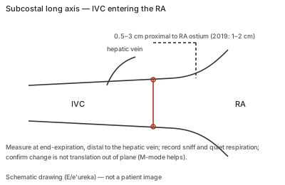
Correct example
Maximum diameter at end-expiration at the stated site, then respirophasic change with quiet breathing and a sniff, confirming the vessel stays in plane; RAP is assigned as 3, 8 or 15 mmHg from diameter and collapse.
Common error
Measuring at the RA junction or over the hepatic vein inflow, measuring an oblique cut, or reading translation of the IVC out of plane during inspiration as collapse.
Effect on interpretation
RAP is added directly to the TR gradient, so a wrong IVC category shifts RVSP by 5–12 mmHg; in positive-pressure ventilation collapse cannot be used and the estimate is indeterminate rather than normal.
Why
The IVC diameter and its collapse with sniff together estimate RAP; indeterminate combinations (normal size without collapse, dilated with collapse) take the intermediate value with secondary indices rather than a guess.