
Echocardiographic screening for suspected pulmonary hypertension
Right heart and pulmonary pressures
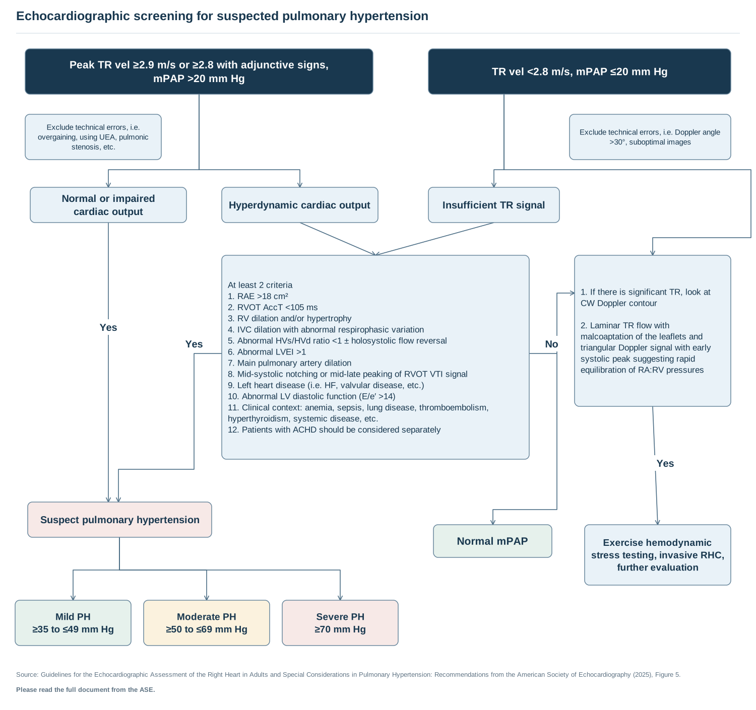
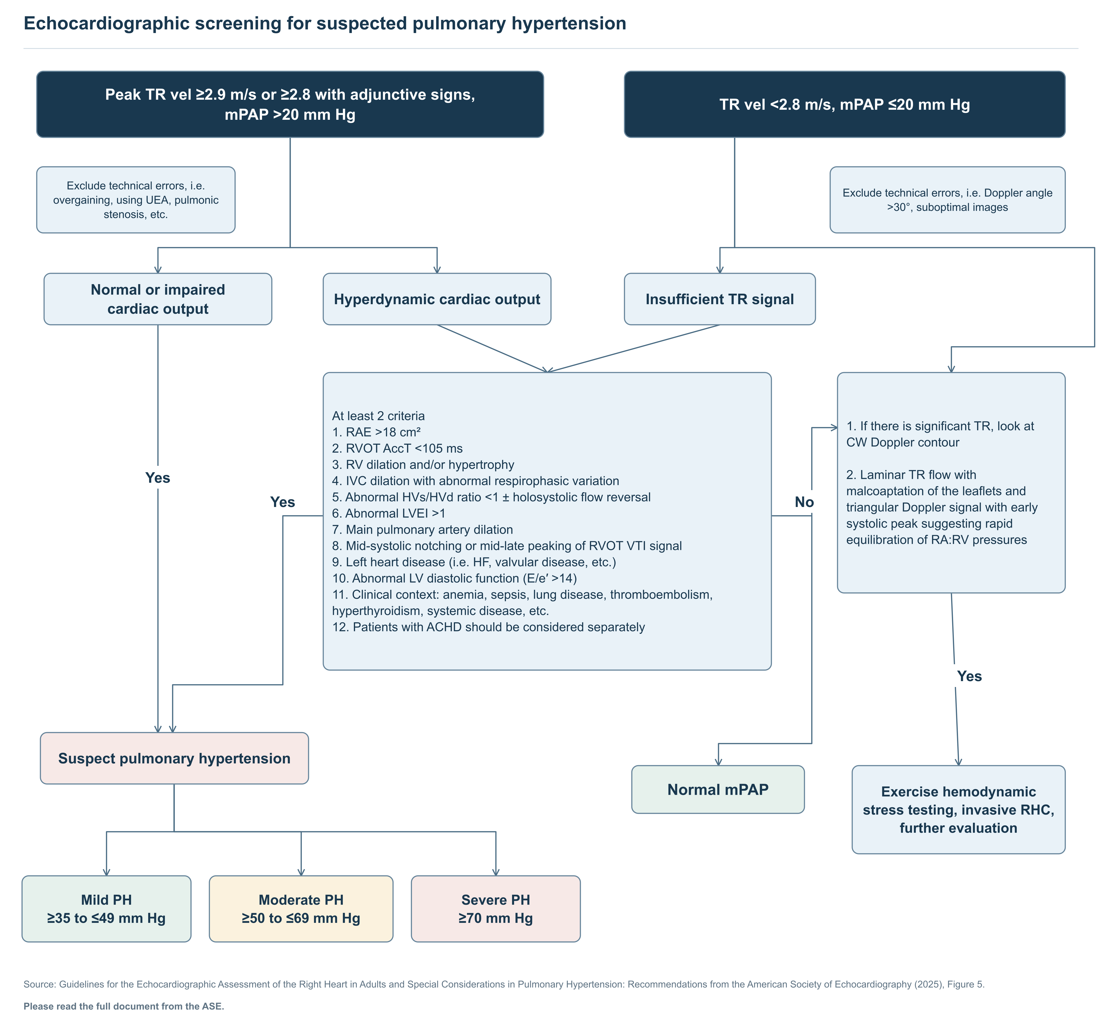
Text of this figure
- Echocardiographic screening for suspected pulmonary hypertension
- No
- Yes
- Yes
- Yes
- Peak TR vel ≥2.9 m/s or ≥2.8 with adjunctive signs,
- mPAP >20 mm Hg
- TR vel <2.8 m/s, mPAP ≤20 mm Hg
- Exclude technical errors, i.e.
- overgaining, using UEA, pulmonic
- stenosis, etc.
- Exclude technical errors, i.e. Doppler angle
- >30°, suboptimal images
- Normal or impaired
- cardiac output
- Hyperdynamic cardiac output
- Insufficient TR signal
- At least 2 criteria
- 1. RAE >18 cm²
- 2. RVOT AccT <105 ms
- 3. RV dilation and/or hypertrophy
- 4. IVC dilation with abnormal respirophasic variation
- 5. Abnormal HVs/HVd ratio <1 ± holosystolic flow reversal
- 6. Abnormal LVEI >1
- 7. Main pulmonary artery dilation
- 8. Mid-systolic notching or mid-late peaking of RVOT VTI signal
- 9. Left heart disease (i.e. HF, valvular disease, etc.)
- 10. Abnormal LV diastolic function (E/e′ >14)
- 11. Clinical context: anemia, sepsis, lung disease, thromboembolism,
- hyperthyroidism, systemic disease, etc.
- 12. Patients with ACHD should be considered separately
- 1. If there is significant TR, look at
- CW Doppler contour
- 2. Laminar TR flow with
- malcoaptation of the leaflets and
- triangular Doppler signal with early
- systolic peak suggesting rapid
- equilibration of RA:RV pressures
- Suspect pulmonary hypertension
- Normal mPAP
- Exercise hemodynamic
- stress testing, invasive RHC,
- further evaluation
- Mild PH
- ≥35 to ≤49 mm Hg
- Moderate PH
- ≥50 to ≤69 mm Hg
- Severe PH
- ≥70 mm Hg
- Source: Guidelines for the Echocardiographic Assessment of the Right Heart in Adults and Special Considerations in Pulmonary Hypertension: Recommendations from the American Society of Echocardiography (2025), Figure 5.
- Please read the full document from the ASE.
Redrawn as a flowchart from the published document named in the figure. Read the full document for context and the figures' complete notes.