
Atrial tracing
| View | Dedicated apical four- and two-chamber views repositioned to maximize the base width and long axis of the left atrium. |
|---|---|
| Timing | End-systole, at maximum atrial volume (frame just before mitral opening). |
| Caliper / trace | Trace the LA blood–tissue border; close the contour with a line across the mitral annulus; exclude the appendage and pulmonary vein confluences; length from annular midpoint to the far superior wall in both views (within 5 mm). |
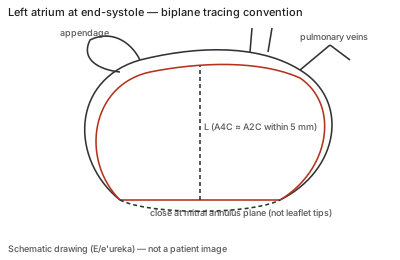
Correct example
A separate atrial acquisition (the optimal LV plane usually foreshortens the atrium), traced at end-systole, closed at the annulus plane rather than the leaflet tips, with the A4C and A2C lengths matching within 5 mm.
Common error
Tracing the atrium on the LV-optimized view, including the appendage or a pulmonary vein in the contour, closing the contour at the leaflet tips, or using views whose long axes differ by more than 5 mm.
Effect on interpretation
Foreshortened atria underestimate volume; included appendage or veins overestimate it. LAVI is a continuous prognostic variable and the 34 mL/m² upper normal limit feeds diastolic grading, so tracing errors propagate into filling-pressure estimates.
Why
The biplane disk summation assumes two orthogonal views through the same long axis; mismatched lengths signal that one view is off-axis, so the images are re-evaluated rather than averaged.