
Steps for diagnosing LV diastolic dysfunction
Atria and diastolic function
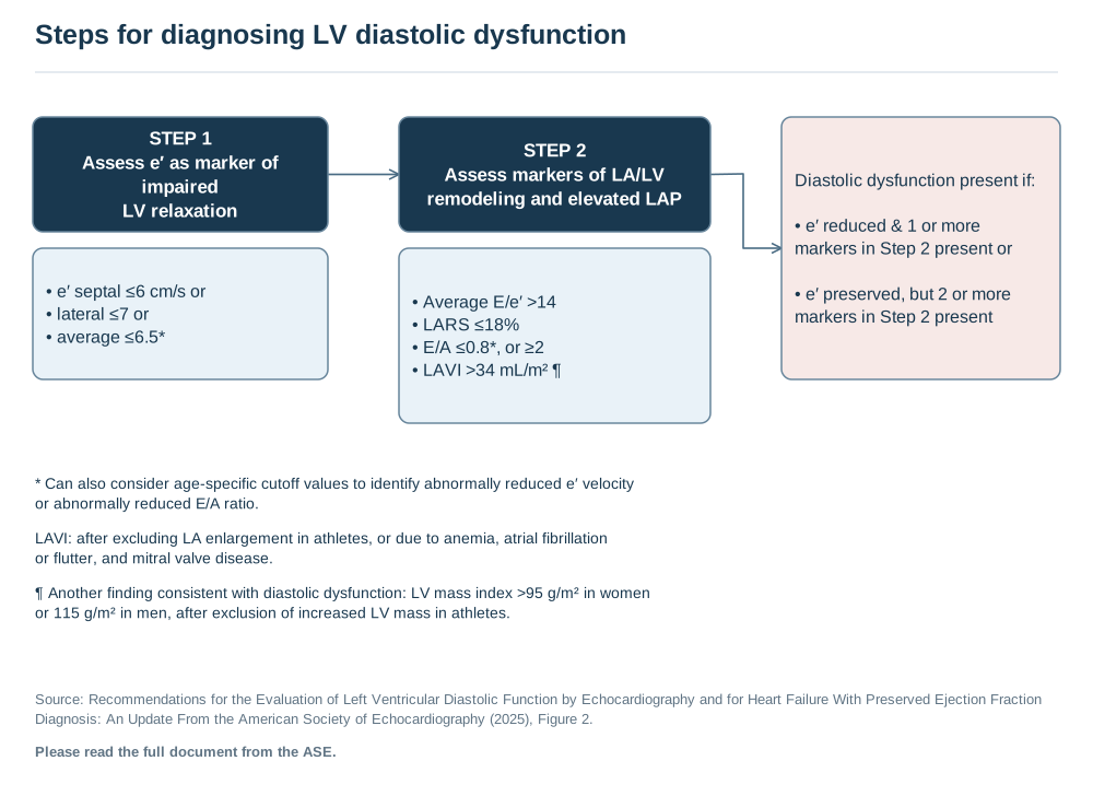
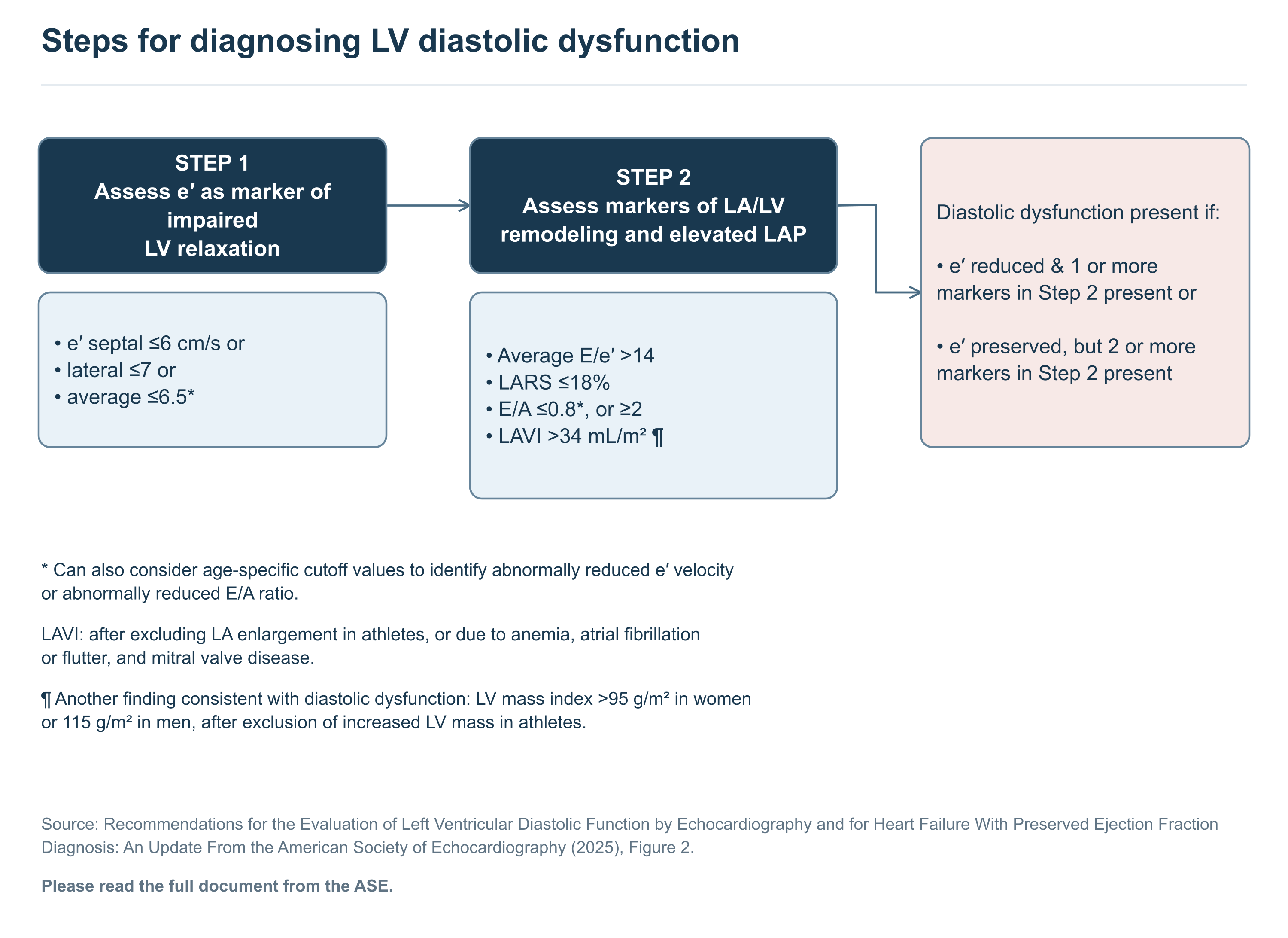
Text of this figure
- Steps for diagnosing LV diastolic dysfunction
- STEP 1
- Assess e′ as marker of
- impaired
- LV relaxation
- • e′ septal ≤6 cm/s or
- • lateral ≤7 or
- • average ≤6.5*
- STEP 2
- Assess markers of LA/LV
- remodeling and elevated LAP
- • Average E/e′ >14
- • LARS ≤18%
- • E/A ≤0.8*, or ≥2
- • LAVI >34 mL/m² 
- Diastolic dysfunction present if:
- • e′ reduced & 1 or more
- markers in Step 2 present or
- • e′ preserved, but 2 or more
- markers in Step 2 present
- * Can also consider age-specific cutoff values to identify abnormally reduced e′ velocity
- or abnormally reduced E/A ratio.
- LAVI: after excluding LA enlargement in athletes, or due to anemia, atrial fibrillation
- or flutter, and mitral valve disease.
- ¶ Another finding consistent with diastolic dysfunction: LV mass index >95 g/m² in women
- or 115 g/m² in men, after exclusion of increased LV mass in athletes.
- Source: Recommendations for the Evaluation of Left Ventricular Diastolic Function by Echocardiography and for Heart Failure With Preserved Ejection Fraction
- Diagnosis: An Update From the American Society of Echocardiography (2025), Figure 2.
- Please read the full document from the ASE.
Redrawn as a flowchart from the published document named in the figure. Read the full document for context and the figures' complete notes.