
LV internal dimension: caliper placement
| View | Parasternal long axis along the centre axis of the ventricle (papillary muscles not visible); biplane imaging can confirm perpendicular alignment. |
|---|---|
| Timing | End-diastole: first frame after mitral closure (or the R-wave peak); end-systole at the smallest cavity, usually the frame before early mitral opening. |
| Caliper / trace | From the compacted septal interface perpendicular to the long axis to the inner border of the compacted posterior wall, just below the mitral leaflet tips; septum and posterior wall at the same level. |
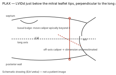
Correct example
Caliper perpendicular to the long axis just beyond the leaflet tips, excluding chordae and papillary apparatus posteriorly and RV trabeculae, moderator band or tricuspid apparatus from the septum; with a basal septal bulge all linear measurements move slightly apical beyond it.
Common error
An oblique caliper (the TTE example shows LVIDd falling from 47 to 44 mm off-axis) or measuring a sigmoid septum at the leaflet tips (LVIDd 38 mm and IVS 17 mm instead of 53 mm and 7 mm beyond the bulge).
Effect on interpretation
Linear LV mass is computed from the cubed difference of these dimensions, so a few millimetres of wall-thickness or cavity error change mass and relative wall thickness categories; EF from linear dimensions is not reported.
Why
The reference ranges were derived from parasternal measurements perpendicular to the long axis at the leaflet-tip level; moving the level or the angle measures a different ventricle than the one the normal values describe.