
Foreshortening
Read more:ASE/EACVI 2015 · Cardiac chamber quantificationASE 2019 · Comprehensive TTE examinationASE/EACVI 2025 · Clinical applications of strain
| View | Apical four- and two-chamber views at reduced depth focused on the LV cavity; dedicated atrial views separately. |
|---|---|
| Timing | Check alignment before tracing end-diastolic and end-systolic frames. |
| Caliper / trace | LV traced along the compacted/noncompacted interface, papillary muscles and trabeculae in the cavity; LV lengths in the two views within 10%. |
Example
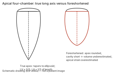
Correct example
Apex at the top centre of the sector, the ventricle tapering to an ellipsoid, and roughly two-thirds LV to one-third LA along the long axis; the transducer is moved laterally or down an interspace until the true apex is seen.
Common error
Accepting a view in which the apex looks rounded and the cavity short; the long axis is cut obliquely and the apex in the image is not the anatomical apex.
Effect on interpretation
Foreshortening underestimates LV volumes and erroneously increases apical segmental strain; it can also hide apical pathology. 3D acquisitions are largely unaffected.
Why
The method of disks assumes the traced contour spans the full long axis; a plane that misses the apex produces a shorter, rounder contour whose summed disks underestimate the true volume.